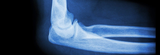
Brachial Plexus Elbow Treatments
Elbow Flexion
The Zancolli procedure does the same trick with the biceps tendon at the elbow. Detached from the radius, it is repositioned on the opposite side to where it was to create pronation. It had produced supination. But what if there is no biceps action at all? Then one can be built. One must work with what is working, as brachial palsies do not pick out single muscles as a rule. For example, the pectoral muscle can be repositioned to become a biceps in certain conditions, but may well be poorly suited in some brachial plexus palsies.
The latissimus dorsi can be detached from the ribs and – pivoting on its attachment at the upper humerus – have the once-rib attachment end attached to the biceps tendon at the elbow. That creates an elbow flexor. That same transfer, if attached to the back of the arm, is used to generate triceps power in conditions wherein triceps substitution is needed.
The finger flexors are in two layers and along with the wrist flexors attach not only in the forearm but also above the elbow at the medial condyle. In a real sense, the finger and wrist flexors are already elbow flexors. They can be recruited as elbow flexors by merely stabilizing the wrist or fingers (as with other muscles). Surgically moving the humeral attachment more proximal increases the leverage of those muscles (which at that level are nearly a single unit). Thus weak elbow flexion can be boosted.
 In the absence of shoulder dislocation, if the external rotation cannot be achieved through the physical therapy program, then surgical release through a subscapularis slide (an advancement of the muscle that blankets the undersurface of the scapula reaching laterally to attaches to the front of the humerus (figure on left)) is performed usually at the age of 18 to 24 months. The child attains, with that, immediate improvement of both function and cosmesis.
If over the course of the ensuing year, the contracture begins to recur, a modified L’Episcopo procedure is performed, where the latissimus and teres major insertions are transferred around to the back of the humerus into the infraspinatus tendon. These muscles are powerful internal rotators of the shoulder that join together in what is called a conjoint tendon which attaches to the front of the arm bone or humerus. This transfer is possible because the shoulder has additional powerful muscles that perform similar movements akin to those of the transferred muscles. This transfer will permanently balance the muscles about the shoulder thus preventing recurrence of the contracture and improving elevation of the shoulder.
 When permanent bony incongruence has developed in the glenohumeral joint, in those patients who are seen late, an external rotation osteotomy is performed to optimize the position of the arc of motion and improve function and appearance (Figure on left). When minimal muscle recovery occurs and there is little or no ability to elevate the shoulder some salvage procedures have been proposed, including shoulder fusion and trapezius transfer. In our center, we believe that shoulder fusion is the last resort. The trapezial transfer leaves the patient with limited abduction, a webbed neck and a poor cosmetic appearance.
The Elbow
In many children with Erb’s palsy, a mild paradoxical elbow flexion contracture often develops. To date, there is no definitive explanation of when and why this occurs. Dr. Price believes it occurs with internal rotation contracture of the shoulder, which tightens the biceps muscle. These mild contractures do not pose any functional problems. It can be reversed in young children by serial casting. If it persists into late childhood, it cannot be reversed without surgery.
Activities of daily living require elbow motion from 30 degrees to 130 degrees. When contractures of the elbow greater than 30 degrees occur, this can not only alter function but also appearance. This severe contracture can occur in a total plexus palsy where there is little or no triceps function, i.e. power to extend the elbow to a straight position. It can also occur when co-contraction exists, involving the biceps and triceps. This is a condition where during the healing of the nerve injury, the “wires get crossed” and elbow flexors will fire while the elbow extensors are trying to extend the elbow. This co-contraction can lead to significant flexion contracture. In small children, this is treated with serial Botox injections and casting. Dr. Price has developed a new operation (which is called the Modified Outerbridge-Kashiwagi Procedure) for older children and adolescents with persistent significant elbow flexion contracture. This procedure involves recontouring the bony elements of the elbow and releasing soft tissue contractures to regain extension.
Elbow flexion nearly always returns, but when the patient lacks the ability to flex the elbow against gravity, a significant functional impairment exists; the patient cannot bring the hand to the mouth for feeding, buttoning a shirt, and combing the hair. Muscle transfers when neurologic recovery has plateaued will give the patient better ability to use the hand. If minor weakness is present, a Steindler flexorplasty is performed [where the origin of the flexor wad of the forearm is moved proximally and centrally to assist in elbow flexion]. If more profound elbow weakness is present, several procedures have been in existence to gain function. There are other transfers possible to gain flexion.
When significant imbalances of the muscles that rotate the forearm are present early in recovery, a pronation (palm down) or supination (palm up) contracture may develop which limits function and sometimes gives an unsightly cosmetic appearance. Even if the opposing muscles recover sufficient power, it, alone, is not capable of reversing a fixed contracture. It is important to release these contractures BEFORE fixed bony deformity occurs in the radius and ulna (the 2 bones of the forearm). When these subtle changes occur, the rotation of the forearm will be permanently restricted. Therefore, a window of opportunity exists in the first few years to release forearm contractures and regain the function of the forearm. If not done early, then rotation osteotomies may be necessary to reorient the arc of motion to a more functional position.
In a classic Erb’s palsy, involvement of the C5 root leads to weakness of the biceps brachii. In addition to being an elbow flexor, the biceps muscle is the most powerful supinator of the forearm, which rotates the palm up. With the significant weakness of the biceps, a pronation contracture may develop which limits function in hygiene, toileting, and sports activities. If the biceps recovers significant power we have demonstrated lasting improvement in function by releasing the pronation contracture in the forearm (ref = Liggio et al).
Conversely, if the biceps recovers enough in a patient with complete plexus injury, the muscle imbalance may cause a supination contracture of the forearm, which is cosmetically unsightly and impairs use of a keyboard, which in today’s computer world is quite problematic. If that occurs and is to be treated before bony changes occur to the radius and ulna, an interosseous membrane release can be performed.
If there is no significant recovery of the pronators of the forearm, which rotate the palm down, a rerouting of the biceps insertion (to reverse the wrap on the radius) will assist in pronating the forearm to neutral pronation (Zancolli procedure). As stated previously, if a treatment is sought late – with contracture and permanent bony changes in the radius and ulna – then rotational osteotomy of these bones to position the forearm in neutral is indicated.
In patients with Total Plexus Palsy or Klumpke’s paralysis, the hand and fingers are affected. The nature of the involvement is highly variable and can include a number of deficits, which require individual assessment for, if necessary, tendon transfers or releases. There are myriad possibilities, too many to generalize. Each child must be individually evaluated and managed with a combination of therapy, splinting and occasionally surgery.
There are facial findings that may be found at birth in cases of brachial plexus injury. Sagging eyelid and pupil size mismatch suggest an additional injury deeper than the plexus itself. Also, note that some embryologic – that is genetic type – disorders of formation can mimic brachial plexus injury. These are related to embryologic gill formation.
Elbow Flexion Contractures in Brachial Plexus Birth Injury Patients
Brachial plexus birth palsy will often lead to elbow flexion contractures. Many of them are minor and require attention in therapy along with splinting or casting to correct or prevent them from worsening. Occasionally, elbow flexion contractures will worsen with the growth and development of the child. There are many theories as to what causes these contractures including muscle imbalance, impaired muscle growth of the elbow flexor muscles, and co-contraction of agonist/antagonist muscles. Until recently, treatment of these contractures was not very effective. With prolonged loss of motion, first, the soft tissues get contracted. Then with growth, the bones making up the elbow joint become permanently deformed. This deformation includes bony overgrowth to form an elongated and broadened of the olecranon.
The fossa or depression at the end of the humerus becomes shallow and too small to accommodate the overgrown olecranon, thus creating a bony block to full extension.
Dr. Price has designed an operation that can permanently and significantly improve the elbow contractures that impair function and alter appearance. Dr. Price has improved and modified the Outerbridge-Kashiwagi operation to benefit patients with Erb’s palsy or brachial plexus birth injury.
The operation narrows and shortens the olecranon, and deepens the olecranon fossa to more closely approximate the normal human anatomy. This is often combined with soft tissue releases in the front of the elbow to restore the ability to more fully extend it.

